The Imaging Center of the Fraunhofer Project Center for Stem Cell Process Engineering offers companies and public institutions a high-grade infrastructure for the analysis of cell processes and cell-material interactions with the aid of live cell imaging. With our extensive range of instruments as well as our materials science expertise, we can support you in sample preparation, planning and execution of test series. We offer measurements to order, carried out by our scientists, as well as rental of equipment, to be used by clients and cooperation partners themselves.
Imaging Center
Fluorescence microscope with time-lapse function and stage top incubator
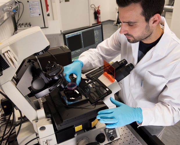
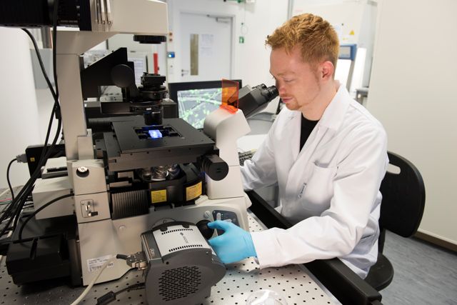
Using the fluorescence microscope, cell processes can be visualized in real time by the stimulation of fluorescent markers. Both, long-term measurements over several hours (e.g. in the area of tissue healing or cell differentiation) as well as the analysis of rapid intercellular processes are possible. Our system allows the implementation of complex measurement sequences including automated multipoint and multichannel measurements of several samples in a single test run. The system is equipped with a stage top incubator to provide the necessary physiological conditions (temperature, CO2 content and air humidity) for all cell types.
Confocal microscope with STED unit
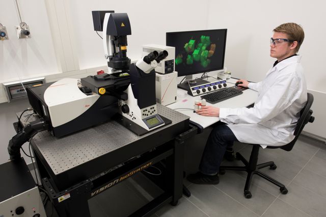
With our confocal STED microscope, fluorescent markers in living cells can be stimulated simultaneously with light of up to eight different wavelengths simultaneously. Due to the special STED technology (STED – Stimulated Emission Depletion) resolutions well below one tenth of the stimulation wavelength can be achieved (super-resolution). Temporally resolved 3D-images can be constructed from the detected fluorescence signal of up to five detection channels in parallel. Single photon detection measurements are also possible.
Spinning disk confocal microscope (SDCM)
The spinning disk confocal microscope enables visualization of the dynamics of living, fluorescence-labelled cells in 3D with an especially high temporal resolution. This allows the examination of cell-surface interactions, the analysis of intracellular processes and the visualization of living cells in three-dimensional scaffolds.
Long time live cell microscopy
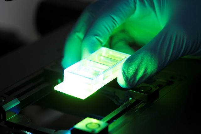
Automated live cell microscopy allows the observation of cellular processes over several days under physiological conditions (37 °C, 5% CO2, > 95% RH). Phase contrast and fluorescence images of several positions in a culture dish can be captured in defined time intervals. The resulting time-lapse recordings combined with automated image analysis supply valuable insights into long time cell-cell and cell-matrix interactions.
 Fraunhofer Project Center for Stem Cell Process Engineering


